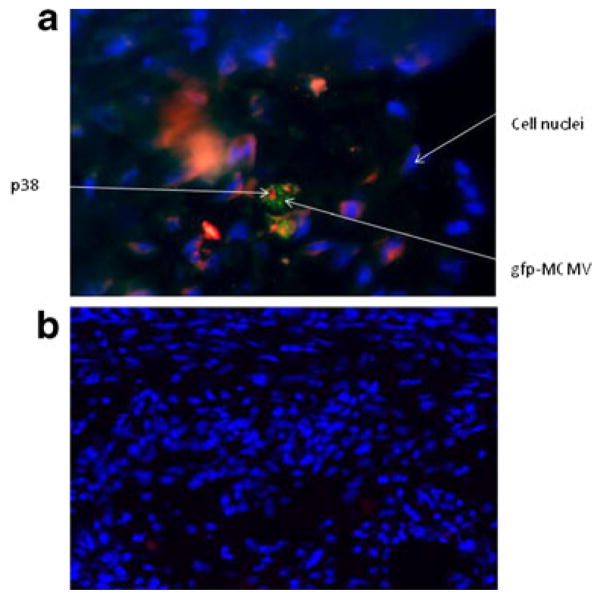
Fig. 5.
Co-localization of gfp-labeled MCMV and p38 in atherosclerotic lesions (figure is representative of eight samples). a Cross-section of an aorta showing an atherosclerotic lesion in a MCMV-infected Apo E KO mouse. MCMV is labeled with gfp (green), p38 with Alexafluor 555 (red), and nuclei with DAPI (blue). Magnification is ×100; b cross-section of an aorta showing an atherosclerotic lesion in an uninfected mouse stained as in (a). Note that only DAPI-stained nuclei are observed (magnification is ×40)

