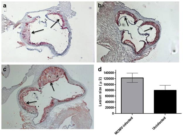
Fig. 6.
Representative figures of atherosclerotic lesions in aortic sinus of MCMV-infected and uninfected Apo E KO mice at 2.5 months post-infection. a Lesions in aortas of uninfected mice (n=8); b lesions in aortas of mice infected with a UV-killed MCMV (n=6; average lesion size 89,928±13,700 μ2; p<0.05 compared to lesions in mice infected with live MCMV); c Lesions in aortas of mice infected with a live MCMV (n=8); d lesion area (μm2±STD) in aortas of MCMV-infected vs. uninfected Apo E KO mice. Values represent average of n=8 mice (MCMV-infected and uninfected), p=0.038. Arrows show atherosclerotic lesions

