
Fig. 4.
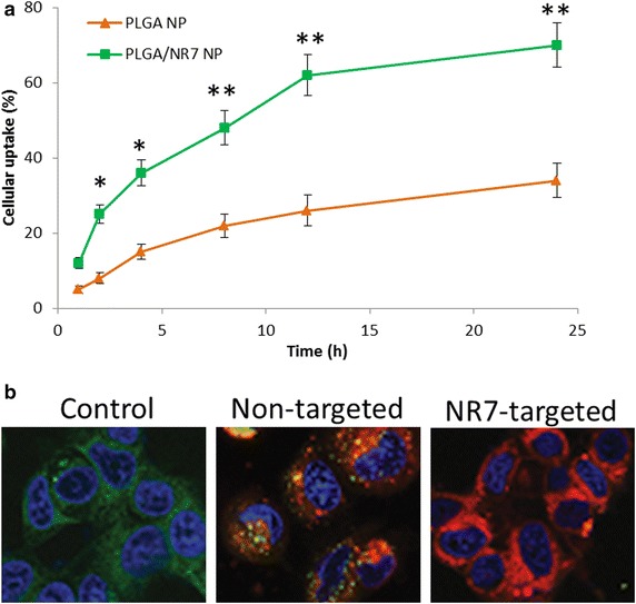
a Intracellular uptake of PLGA NP and PLGA/NR7 NP in HN6 squamous cell carcinoma. Rhodamine B was used as a fluorescent dye. The uptake is shown as a percentage of total amounts of NP (dye) incubated with the cancer cells. b Representative confocal microscopy images of targeted and non-targeted NP in HN6 cancer cells. The cells are stained with Lysotracker lysosomal stain and DAPI was used to stain the nucleus. **p < 0.01 and *p < 005 is the statistical difference between the cellular uptake of two formulations