
Fig. 4. White matter injury in aged mice is linked with functional deficits at late stages of dMCAO.
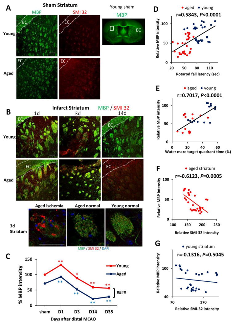
A. MBP and SMI-32 staining in sham-operated young and aged mice. White squares in lower magnification views of MBP staining designate the area shown under high magnification on the left. EC, external capsule. B. Representative images of MBP and SMI-32 double-staining at 1, 3, and 14d after dMCAO in young and aged mice. Scale bar: 100 μm. High power images of the striatum at 3d after dMCAO were shown in lower panels. Scale bar: 50 μm. C. Quantification of MBP staining intensities in the striatum in young and aged mice after sham-operation or at 1, 3, 14, and 35 days after dMCAO. Data were expressed as percentage of young sham. n=7 per group. *p≤0.05, **p≤0.01, vs corresponding sham. ####p≤0.0001 young vs aged. D-E. Pearson correlation between MBP staining intensity and behavioral performance on the Rotarod or Morris water maze memory tests. D. There was a positive correlation between MBP staining intensity and Rotarod performance at 35d after dMCAO. E. There was a positive correlation between MBP intensity and water maze performance at 21d after dMCAO. F-G. Pearson correlation between MBP and SMI-32 staining intensity. F. There was a negative correlation between SMI-32 intensity and MBP intensity in aged mice after dMCAO. G. There was no significant correlation between SMI-32 intensity and MBP intensity in young mice after dMCAO.