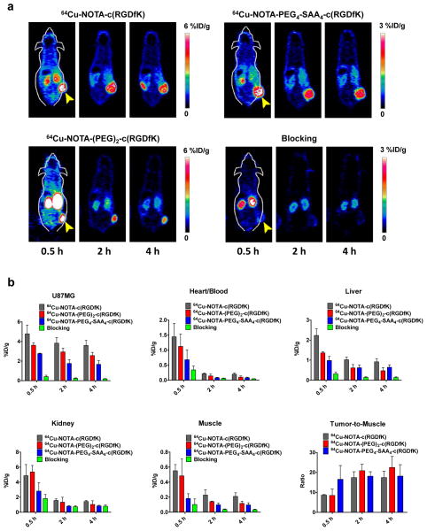
Fig. 4.
Noninvasive microPET imaging of tumor-associated integrin αvβ3 in athymic nude mice bearing U87MG tumors. (a) Representative coronal PET images of planes containing U87MG tumors, at 30 min, 2, and 4 h after intravenous injection of 5.5 MBq of 64Cu-NOTA-c(RGDfK), 64Cu-NOTA-(PEG)2-c(RGDfK), 64Cu-NOTA-PEG4-SAA4-c(RGDfK), or 64Cu-NOTA-PEG4-SAA4-c(RGDfK) coinjected with an c(RGDyK) (10mg/kg) blocking dose; yellow arrow heads indicate the location of the tumor. (b) Quantitative analysis of the PET images showing the timecourse of the accumulation of the tracers in U87MG tumor, blood pool, liver, kidneys, and muscle. Uptake values are expressed as %ID/g ± SD (n = 3). Bottom right panel describes the tumor-to-muscle ratios attained with each of the radiolabeled peptides.

