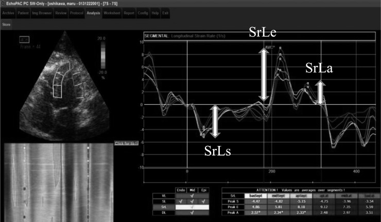
Fig. 1.
Longitudinal strain rate tracking for one cardiac cycle obtained from the left 4-chamber apical view. Six curves with different colors depict respective myocardial segments of left ventricle (basal, middle and apical regions of the septal and lateral). Systolic and early diastolic values of longitudinal strain rate (SrLs, SrLe and SrLa) were calculated.

