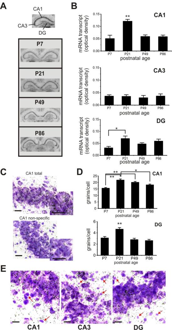
Fig. 3.
Postnatal expression of Zfp804a mRNA varies in hippocampal areas across postnatal life. (A) Representative images of coronal sections showing the hippocampus at different postnatal days. (B) Top, Zfp804a gene expression within area CA1 peaks at P21, which is significantly higher than other time points (Tukey's HSD, P<0.005). Middle, gene expression is fairly low and stable throughout postnatal life in area CA3. Bottom, within the DG, Zfp804a gene expression is significantly lower at P7 compared to P21 (Tukey's HSD, P<0.05), but does not differ from other time points. (C) Representative examples of total and non-specific silver grain labeling within CA1. Dark silver grains show cellular localization of Zfp804a mRNA within thionin-stained hippocampal neurons. Insets show magnified view of labeled neurons. (D) Silver grain density for CA1 and DG confirmed the pattern of elevated mRNA at P21 in both hippocampal regions. (Tukey's HSD, * P<0.001, ** P<0.0005) (E) A subset of smaller, thionin-positive cells that are morphologically consistent with glial cells, show a marked absence of silver grain labeling (red arrows) in all three hipppocampal regions. All scale bars: 10 μm.

