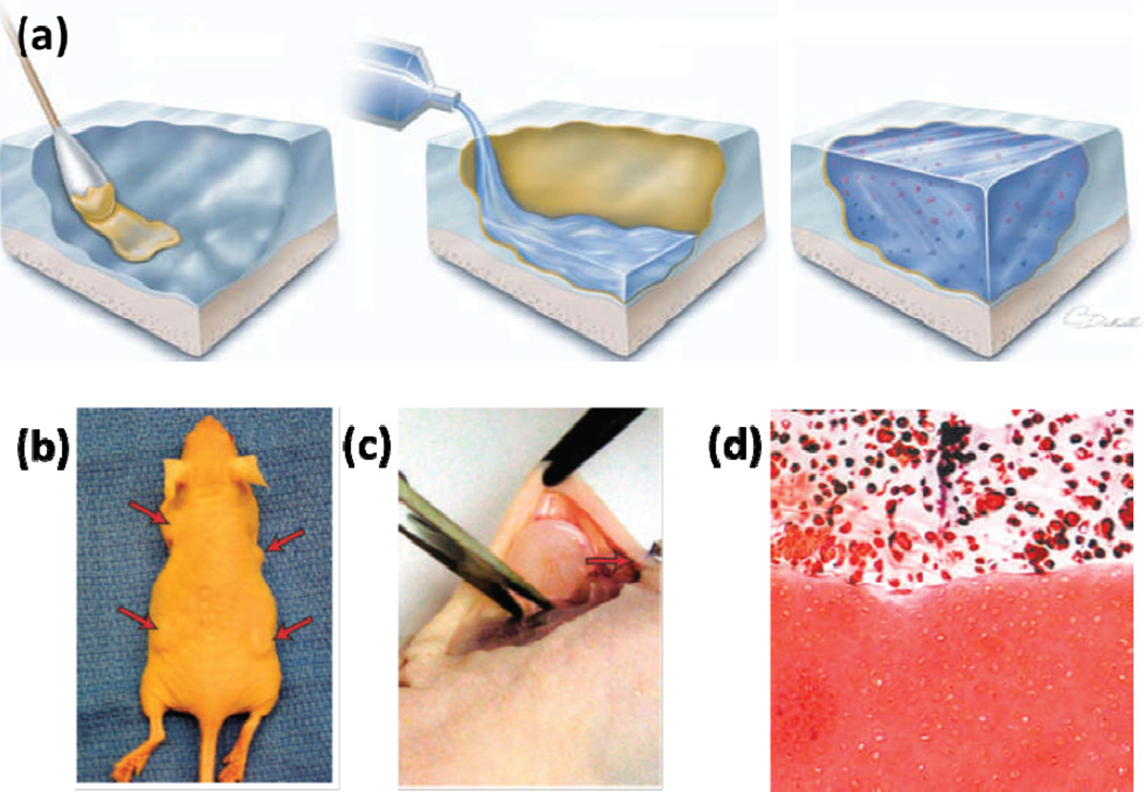
Figure 4.
(A) Schematic diagram showing application of the chondroitin sulfate-based adhesive for hydrogel-tissue integration. The yellow colored layer indicated the chondroitin sulfate layer which served as the bridge between the cartilage tissue and the hydrogel. (B) In vivo subcutaneous implantation of the integrated cartilage-hydrogel constructs in a mice model. (C) Sample explantation after 5 weeks. (D) Safranin-O was found throughout the hydrogel layer and at the interface between the engineered and native cartilage tissues. Adapted from Ref [66] with permission from Nature Publishing Group, copyright 2007.

