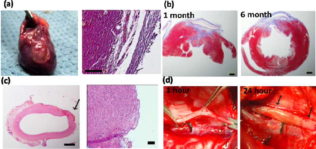
Figure 5.
Highly elastic PGS-based glue for cardiovascular surgeries. (a) Explanted rat heart treated by the engineered glue after 14 days and corresponding H&E staining of the tissue in contact with the glue. (b) H&E and MT staining of the rat cardiac tissue after 1 and 6 months of defect closure with the glue, showing the formation of scarring and accumulation of organized collagen (scale bars: 1mm). (c) H&E staining of Pig carotid artery after treating with glue (scale bars: 1 mm (left) and 50 µm (right). The arrow points to the defect created. (d) Pig carotid artery one hour after incision creation and 24 hours after closure with HLAA. No bleeding was detected at the defects after 24 h of operation, as indicated by arrows. Adapted from Ref [5] with permission from the AAAS, copyright 2014.

