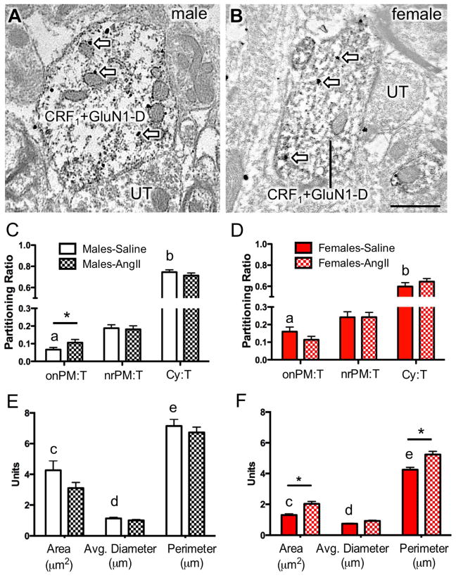
Figure 2. Dual GluN1 and CRF1-EGFP containing dendrites of the RVLM in male and female mice administered saline or AngII: Subcellular distribution of GluN1 and dendritic morphology.
(A, B) Representative images of dendrites from male (A) and female (B) mice containing both diffuse immunoperoxidase labeling for CRF1-EGFP and black particulate labeling for GluN1 (arrows). (C, D) The partitioning ratio is shown as the number of SIG particles on the plasma membrane (onPM), near the plasma membrane (nrPM) or in the cytoplasm (Cy) relative to the total number of particles (T). In the absence of AngII, CRF1-EGFP dendritic profiles from females show a significantly higher ratio of GluN1-SIG on the plasma membrane (onPM) compared to CRF1-EGFP dendritic profiles from males. Following AngII, male mice show a significant increase in the ratio of onPM GluN1-SIG labeling. (E, F) In the absence of AngII, the area and the perimeter of CRF1-EGFP containing dendrites is significantly larger in males compared to females. Following AngII, the area of CRF1-EGFP dendritic profiles is significantly higher in AngII females compared to saline-treated females. *: P < 0.05 in saline versus AngII treated mice. a: P < 0.05 in female versus male mice given saline; b,c,d: P < 0.05 in male versus female mice given saline. UT, unlabeled terminal. Scale bar = 500 nm

