
Figure 9.
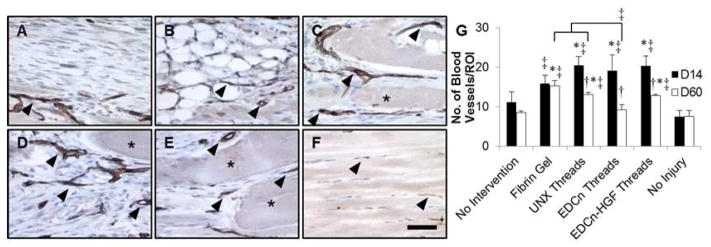
IHC staining of PECAM in VML defects 14 days post-injury. IHC staining for PECAM with a hemotoxylin counterstain of VML defects with (A) no intervention, (B) fibrin gel, (C) UNX microthreads, (D) EDCn microthreads, (E) EDCn-HGF microthreads, or (F) uninjured muscle. Asterisks indicate microthreads and black arrowheads denote representative blood vessels. Scale = 50 μm. (G) Quantification of the number of PECAM positive blood vessels in VML defects. ‡ and brackets indicate significant differences between indicated groups as determined by one-way ANOVA with Holm-Sidak post hoc analysis, † denote significance with D14 values as determined by Student’s t-test (p<0.05, n=3 for D14, n=6 for D60).