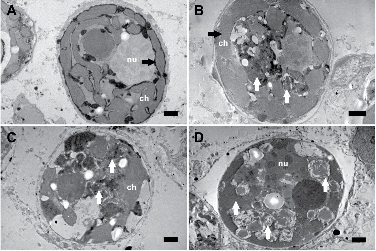
Fig 3. Representative transmission electron micrographs documenting the effects of thermal stress on the internal structure of endosymbiotic Symbiodinium cells within tissue of Acropora millepora.
(A) Symbiodinium exposed to 27°C showing intact organelles and thylakoid membranes (black arrow). (B) First signs of degraded internal structures in some Symbiodinium cells after 7 days of heat stress (white arrows). Note the intact structure of the thylakoid membranes (black arrow). (C and D) Symbiodinium exposed to 32°C revealing degraded internal structures (white arrows). Scale bars, 1 μm; ch, chloroplast; nu, nucleus.

