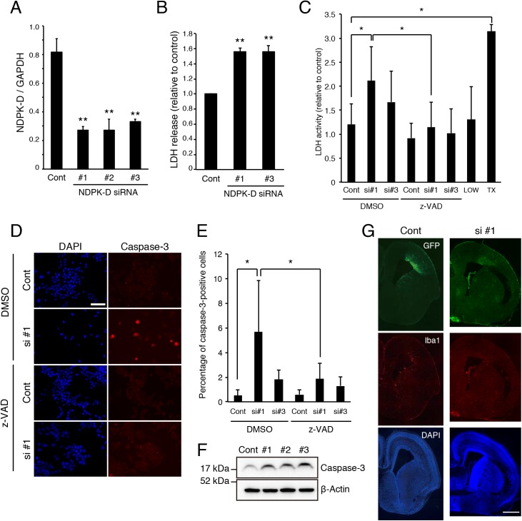
Fig 2. NDPK-D Knockdown induces apoptosis of N1E-115 cells.
(A) NDPK-D siRNAs reduced NDPK-D mRNA expression. N1E-115 cells were transfected with the indicated siRNAs. Total RNA isolated at 72 h post-transfection was analyzed by real-time PCR. **P < 0.01. n = 3. (B) siRNA-mediated knockdown of NDPK-D increased LDH release. N1E-115 cells were transfected with indicated siRNA and cultured for 48 h. The relative LDH activities were measured in the culture medium and normalized with control values. **P < 0.01. n = 3. (C) z-VAD-FMK rescues NDPK-D siRNA induced apoptosis. N1E-115 cells were transfected with indicated siRNA and treated with or without 50 μM z-VAD-fmk. Cells were cultured for 48 h and LDH activities were measured as described in (B). LOW: no transfection; TX: 0.1% triton-X 100; *P < 0.05. n = 3. (D, E) siRNA-mediated NDPK-D knockdown increased the number of cleaved caspase-3-positive cells. N1E-115 cells transfected with indicated siRNAs were immunostained with anti-cleaved caspase-3 antibody. The representative images of transfected N1E-115 were shown (D). Percentage of cleaved caspase-3-positive cells were demonstrated in the graph (E). *P < 0.05. Scale bar: 100 μm. n = 3. (F) NDPK-D knockdown produces cleaved caspase-3. N1E-115 cells were transfected with indicated siRNAs. Cell lysates were prepared 72 h after transfection and subjected to western blotting. Cont: control siRNA; si #1, 2, 3, NDPK-D siRNA #1, 2, 3. (G) Knockdown of NDPK-D increased cell death in mouse embryo. Representative images of E17 brain sections from embryos that were co-transfected with GFP and NDPK-D siRNA #1 at E14. The sections were immunostained with anti-GFP and anti-Iba1 antibodies. Scale bar: 600 μm. Statistical analyses were performed using one-way ANOVA followed by Scheffe’s (A, B), or Tukey-Kramer’s (C, E) multiple comparison tests.

