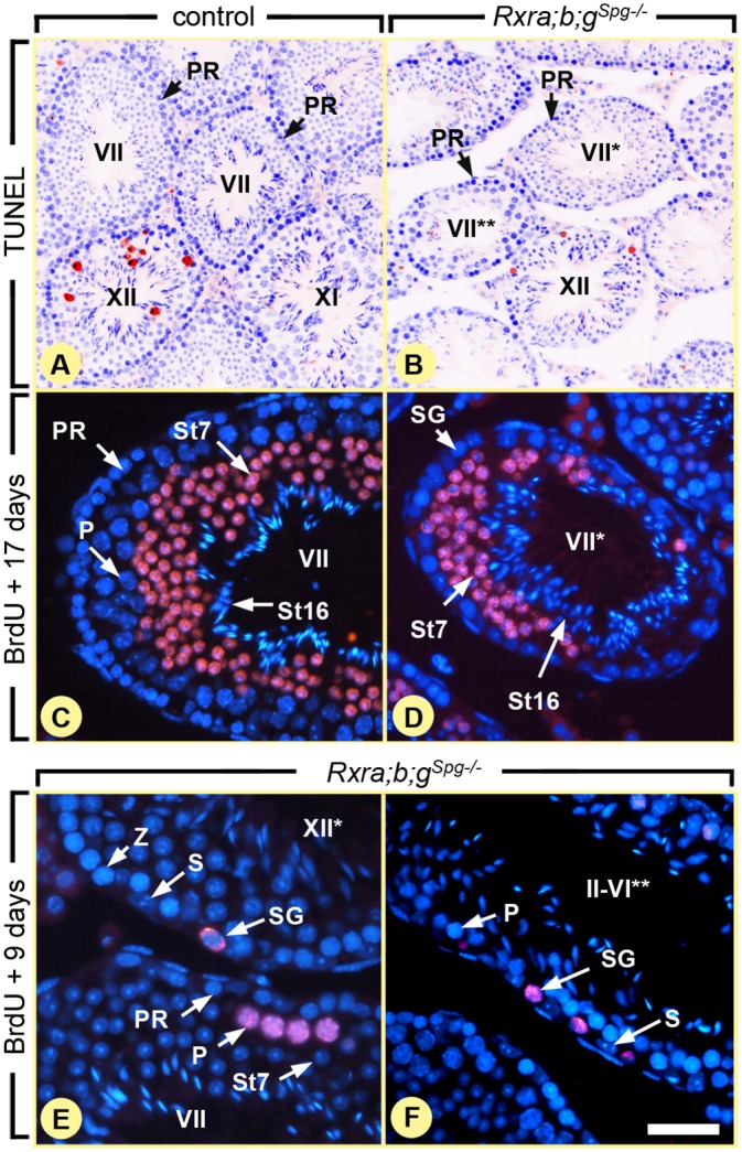
Fig 4. Ablation of RXR in spermatogonia blocks their division, but does not affect meiosis.

(A,B) TUNEL assays on histological sections from 8 week-old control and Rxra;b;g Spg–/– testis as indicated. Red signals correspond to apoptotic cells and nuclei are counterstained with DAPI (in blue). (C-F) Immunohistochemical detection of BrdU (red signals). After administration, incorporated BrdU has been similarly transferred to spermatids at 17 days (C,D) or to pachytene spermatocytes at 9 days (E,F) both in control and mutant seminiferous tubules. In contrast, spermatogonia retaining BrdU are observed only in mutants (E,F). PR and P, preleptotene and pachytene spermatocytes, respectively; S, Sertoli cells; SG, spermatogonia; St7 and St16, step 7 and 16 spermatids, respectively; Z, zygotene spermatocytes. Roman numerals refer to the stages of the seminiferous epithelium cycle. In mutant testes, one asterisk and two asterisks indicate tubule sections without pachytene spermatocytes and without round spermatids, respectively. Scale bar: 160 μm (A,B), 40 μm (C,D) and 25 μm (E,F).
