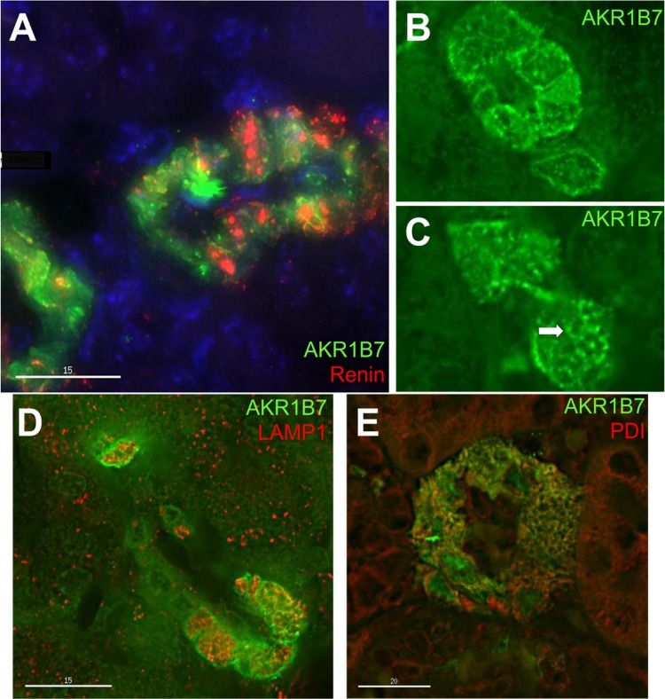
Fig. 3.
Confocal immunofluorescence for AKR1B7, renin, and organelle markers in JG cells. A: immunofluorescence for AKR1B7 (green) and renin (red), with counterstaining with DAPI (blue). The two proteins are found in the same cells, but seldom coincide subcellularly (yellow). B and C: immunofluorescence for AKR1B7 (green). AKR1B7 can be found in or near the plasma membrane (B), and also in lattice-shaped structures in the cytoplasm (C, arrow). D: Immunostaining for AKR1B7 (green) and LAMP1 (red), a marker for lysosomes. The two only show occasional colocalization. E: AKR1B7 (green) and protein disulfide isomerase (red), a marker for endoplasmic reticulum. The two show extensive colocalization (yellow).

