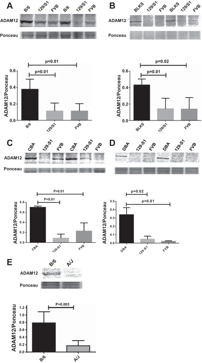
Fig. 3.
Mouse strains with better perfusion recovery (C57Bl/6, C57BlKS, and CBA) show higher ADAM12 protein expression compared with strains with poor perfusion recovery (129-S1, FVB, and A/J). Representative blots showing higher expression of ADAM12 in wk 5 postischemic hindlimb muscles (gastrocnemius) of C57Bl/6 (B/6) compared with 129-S1 and FVB (A); C57BlKS compared with 129-S1 and FVB (B); CBA compared with 129-S1 and FVB (C); DBA compared with 129-S1 and FVB (D); and C57Bl/6 compared with A/J (E). E, top: ADAM12 Western and Ponceau staining; bottom: quantification of bands in top relative to same region on the Ponceau-stained blot (n = 3–4/group).

