Fig. 6.
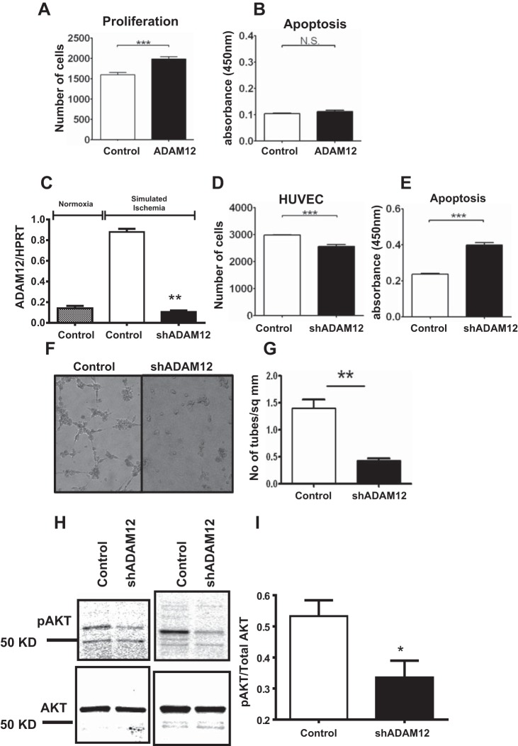
Augmentation of ADAM12 increased EC proliferation, whereas knockdown of ADAM12 decreased EC proliferation, survival, angiogenesis, and AKT phosphorylation (p-AKT) in simulated ischemia. A: human ADAM12 cDNA-transfected HUVECs show increased proliferation and no change in apoptosis (B; proliferation and apoptosis assays, n = 6–10/group, ***P < 0.01, NS = P > 0.05). C: transfection of shRNA targeting human ADAM12 results in a significant decrease in ADAM12 mRNA expression (n = 6/group, **P < 0.01). D: shADAM12-transfected HUVECs show decreased proliferation and increased apoptosis in simulated ischemia (E; proliferation and apoptosis assays, n = 6–10/group, ***P < 0.01). F: representative picture showing that shADAM12-transfected HUVECs have decreased tube formation. G: assessment of the number of tubes formed by shADAM12-transfected HUVECs compared with HUVECs transfected with control plasmid (n = 6/group, **P < 0.01). H: representative picture of Western blot from shADAM12-transfected HUVECs probed with anti-AKT and anti-p-AKT. I: quantification of blot in H showing p-AKT:AKT ratio (n = 5/group, *P < 0.05).

