
Fig. 3.
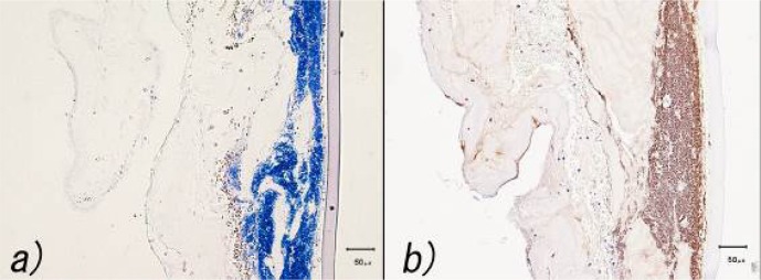
Histopathological examination of the corneal lesion. a) Gram staining demonstrated Gram-positive cocci. Hemorrhage and infiltration of polymorphonuclear neutrophils were observed in the stroma. b) Immunohistochemical staining with a monoclonal anti-Staphylococcus aureus antibody revealed Gram-positive cocci and the cytoplasm of the polymorphonuclear neutrophils to be positive.