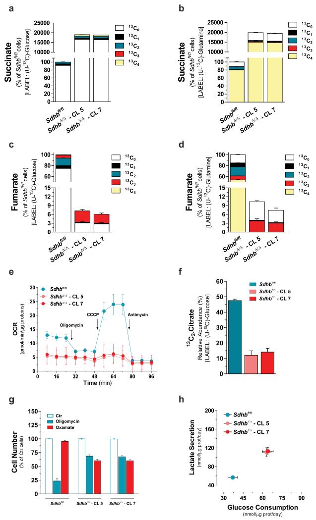
Figure 1. Loss of Sdhb is sufficient to truncate TCA cycle and disable mitochondrial respiration.
Isotopologues distribution of intracellular succinate and fumarate after incubation for 24h with either U-13C-glucose (a, c) or U-13C-glutamine (b, d). All data are presented as mean±s.e.m of one representative experiment (n=3 wells) independently replicated twice (c, d) and three times (a, b). (e) Respiratory profile of the specified cells. Arrows indicate incubation of cells with the indicated compounds. Data are presented as mean±s.e.m of n=27 (Sdhbfl/fl), n=31 (SdhbΔ/Δ – CL 5) and n=29 (SdhbΔ/Δ – CL 7) wells pooled from four independent experiments. (f) Fraction of intracellular citrate pool containing two 13C atoms as result of pyruvate dehydrogenase activity. Data are presented as mean±s.e.m of one representative experiment (n=3 wells) independently replicated twice. (g) Cell number of the indicated cells measured after 96h of treatment with 0.5 μM oligomycin or 5 mM oxamate. Data are presented as mean±s.e.m of n=12 wells pooled from four independent experiments. (h) Rates of the indicated metabolites upon 48h of incubation of Sdhbfl/fl and SdhbΔ/Δ cells with U-13C-glucose. The sum of all isotopologues is reported for clarity. Data are presented as mean±s.e.m of n=18 wells pooled from three independent experiments. In this and all other figures, “wells” represent technical replicate samples set up and assessed under identical conditions and in parallel within a single experiment. Raw data of independently repeated experiments are provided in Supplementary Table 3.

