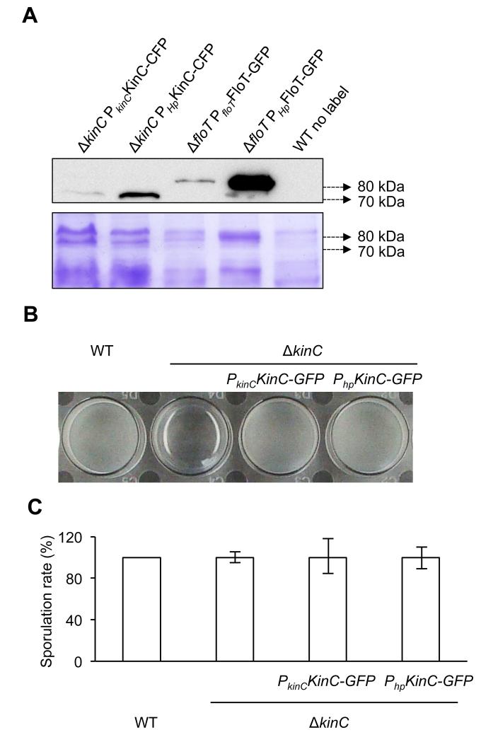
Figure 1. Overexpression of physiologically relevant levels of KinC.
(a) Immunoblot detection of native and induced levels of KinC-CFP (left lanes) and FloT-GFP (right lanes) in B. subtilis. Native promoters are represented by PkinC and PfloT, respectively. IPTG-inducible promoter is represented by Php. Right lane is unlabeled wildtype strain (WT) that served as negative control. SDS-PAGE is shown as the loading control. (b) Pellicle formation assay in different genetic backgrounds. KinC-deficient strain is unable to form pellicles. Complementation of the ΔkinC mutant with KinC-GFP translational fusion recovered the ability to form pellicles to wild type levels. Expression of KinC-GFP under the native or IPTG-induced promoter does not affect pellicle formation. Pellicle formation assay was performed in MSgg medium. Cultures were allowed to grow at 30°C overnight. A more detailed protocol for the pellicle formation assay is described in figure S4. (c) Sporulation rate in ΔkinC mutant and complemented strains is similar to WT levels. Cultures were grown in MSgg medium at 30°C overnight.

