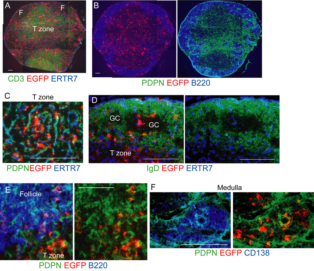
Figure 1. Non-T non-B CD11c+ cells localize with PDPN+ cells in multiple compartments.
Cd11c−DTRRag1−/− mixed chimeras were immunized in footpads with OVA-Alum at day 0 and draining popliteal nodes were taken at day 9. Sections were stained for the DTR-EGFP fusion protein and other indicated markers. (A–B) Nearby sections from the same lymph node showing EGFP+ cell localization relative to (A) T cells and (B) B cells and PDPN+ cells. F=follicle. (C–F) EGFP+ cell localization in the (C) T zone (D) follicles, (GC=germinal center), (E) follicular mantle zone, (F) medulla. Results representative of at least 3 lymph nodes. For (A-B, D, F), bar = 100um. For (C,E), bar=50um. See also Figures S1-S2.

