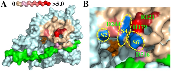
Figure 4.
Binding epitope of mAb 1B10.1 on BoNT/B surface. Shown are molecular models constructed with Pymol software based on the BoNT/B crystal structure (pdb ID: 1S0F). (A) Holotoxin LC structure with color-coding indicating the change in ΔΔG (kcal/mole) of binding of 1B10.1 using the scale shown at top left. The residues comprising the active site are shown in orange; (B) Expanded view of the 1B10.1 epitope on the surface of BoNT/B with color-coding as in (A). Substrate-binding S-pockets are shown in blue. The 1B10.1 epitope includes the S2 amino acid D244 and would cover the S4 and S6 binding sites of Syb-2.

