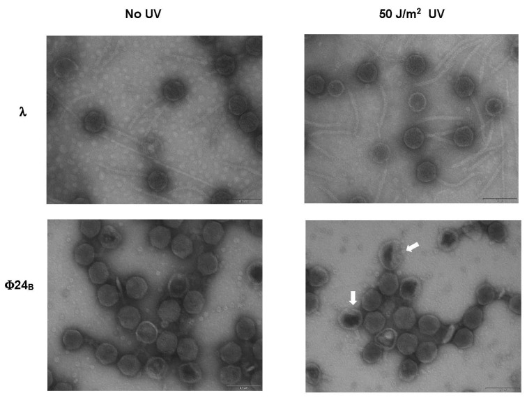
Figure 3.
Electron micrographs of virions of bacteriophage λ (upper panels) and Stx phage Φ24B (lower panels), either non-irradiated (left panels) or irradiated with UV light at 50 J/m2 (right panels). Untypical Φ24B virions with partially damaged heads are indicated by arrows in the lower right panel. Bars, shown at the lower right corner of each panel, correspond to 100 nm.

