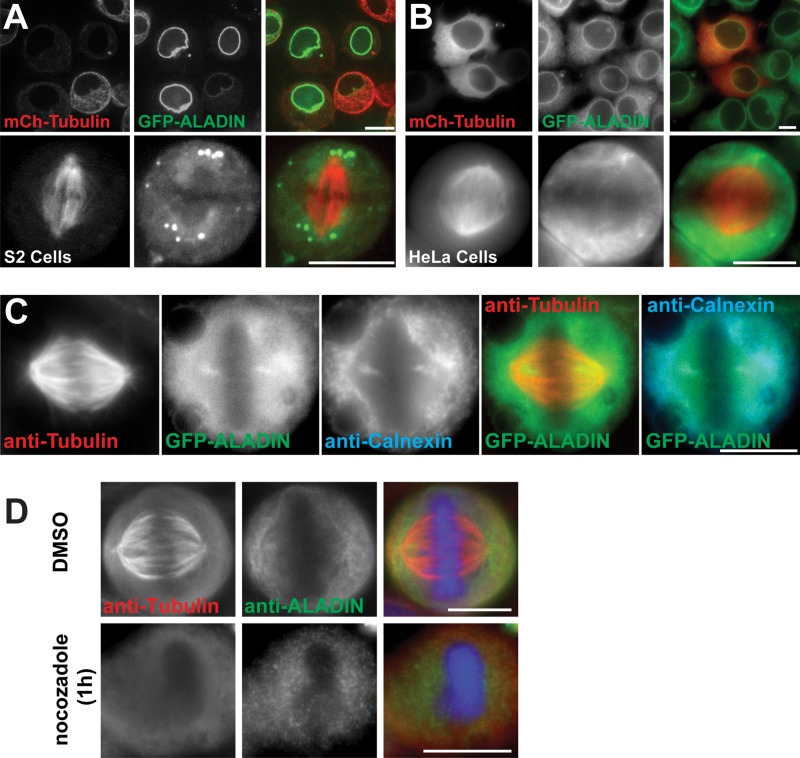
FIGURE 2:
ALADIN localizes around the mitotic spindle and at the spindle poles in Drosophila and human cells. (A) Drosophila S2 cells expressing GFP-ALADIN and mCherry–α-tubulin in interphase (top) or metaphase (bottom). (B) Representative images of interphase (top) and metaphase (bottom) HeLa cells stably expressing GFP-ALADIN and transiently transfected with mCherry–α-tubulin. (C) GFP-ALADIN partially colocalizes with calnexin, an integral ER protein. (D) HeLa cells were fixed in the presence or absence of nocodazole and then stained to visualize tubulin and ALADIN. Scale bars, 10 μm.

