
FIGURE 3:
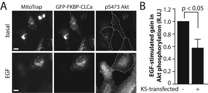
Knocksideways silencing of clathrin light chain reduces EGF-stimulated Akt phosphorylation in ARPE-19 cells. ARPE-19 cells were cotransfected with cDNA encoding MitoTrap (pMito-mCherry-FRB) or GFP-FKBP-CLCa, after which cells were treated with 1 μM rapamycin for 10 min or left untreated (Supplemental Figure S3) and then stimulated with 5 ng/ml EGF for an additional 5 min in the continued presence of rapamycin. After this, cells were fixed and processed for immunofluorescence staining using anti–phospho-Akt (pS473)-specific antibodies. (A) Representative micrographs; scale bar, 20 μm. (B) Mean fluorescence intensity of phospho-Akt (pS473) staining was quantified in cells expressing both MitoTrap and GFP-FKBP-CLCa (KS transfected +) or only one of these exogenous fluorescent proteins (KS transfected –). Mean ± SE of EGF-stimulated gain in phospho-Akt (pS473) levels (n = 3).