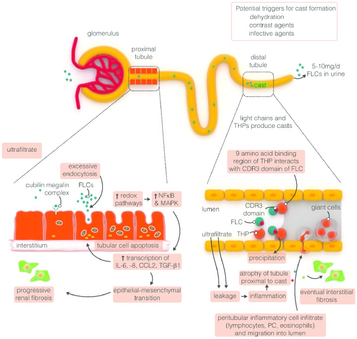
Figure 2.

Mechanisms of FLC-induced acute kidney injury. Serum FLCs are primarily cleared by the kidneys through glomerular filtration, endocytosed by the proximal tubule cells and degraded within lysosomes.50 In MM, the Ig light chains are produced in excess and absorption mechanisms in the proximal tubule are overwhelmed. Thus, the excessive light chains reach the distal tubules, where they form tubular casts with Tamm-Horsfall protein (THP), subsequently leading to tubular obstruction.51 Additionally, excess FLCs can cause direct injury to proximal tubular cells through the induction of pro-inflammatory cytokine production and other pathways leading to tubular cell death.52 The very high concentrations of FLCs present in the ultrafiltrate of patients with MM can result in direct injury to PTCs. Activation of redox pathways occurs, with increased expression of NFκB and MAPK, which in turn leads to the transcription of both inflammatory and profibrotic cytokine. In the distal tubules, FLCs can bind to a specific binding domain on THPs and co-precipitate to form casts. These casts result in tubular atrophy proximal to the cast and lead to progressive interstitial inflammation and fibrosis. CCL2: hemokine (C-C motif) ligand 2; CDR: complementarity determining region; FLC: free light chain; IL: interleukin; MAPK: mitogen-activated protein kinase; NFκB: nuclear factor κB; PTC: proximal tubule kidneys; TGF-β1: transforming growth factor β1; THP: Tamm-Horsfall protein (adapted to Hutchison et al.49).
