Figure 4.

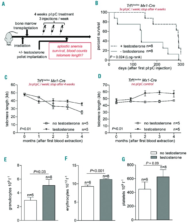
In vivo androgen therapy during moderate Trf1 deletion. (A) Experimental design. Eight-week old wild-type mice were transplanted with bone marrow (Trf1lox/lox Mx1-Cre). After a latency period of 1 month Trf1 deletion was induced by three pI:pC injections per week for a total of 4 weeks. Mice were then subcutaneously implanted with a slow-release testosterone pellet. (B) Kaplan-Meier survival curve of mice treated as detailed above. A log-rank (Mantel-Cox) test was used for statistical analysis. The P-value is depicted. (C) High throughput-Q-FISH longitudinal analysis of telomere length in peripheral blood leukocytes in mice treated with or without testosterone. All mice also received pI:pC injections three times per week for a total period of 4 weeks. (D) High throughput-Q-FISH longitudinal analysis of telomere length in peripheral blood leukocytes in mice not treated with pI:pC and treated or not with testosterone. (E) Granulocyte, (F) erythrocyte and (G) platelet counts 3 months after pI:pC treatment was stopped. Mice given or not given androgen therapy are compared. All graphs show mean values, error bars indicate SEM, n = number of mice. A two-way ANOVA test was used for statistical analysis in (C) and (D); a two-sided Student t-test was used for the experiments shown in (E–G). P-values are indicated.
