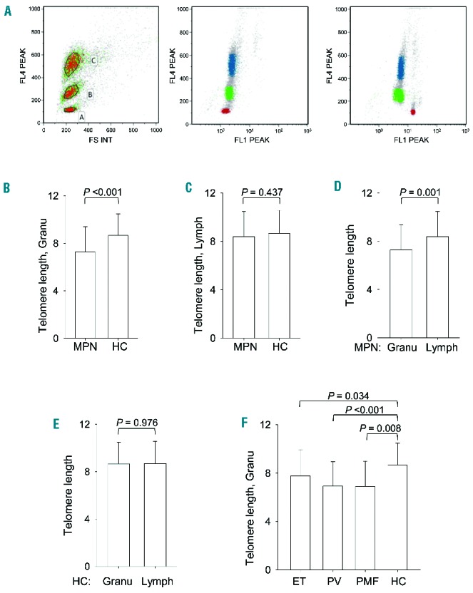
Figure 1.

Flow-FISH assessment of telomere length in blood cells from MPN patients. (A) (Left) Forward scatter on the x-axis and cell cycle stain with LDS 751dsDNS staining, detectable in FL4, on the y-axis. Cell populations were gated as shown, where gate A includes calf thymocytes (red in middle and right panels), gate B human mononuclear cells (green in middle and right panels) and gate C human granulocytes (blue in middle and right panels). (Middle) Whole blood sample hybridized without telomeric probe. (Right) Whole blood sample hybridized with FITC-labeled telomeric probe. (B–F) Telomere length (kB) in granulocytes (Granu) and lymphoid cells (Lymph) derived from patients with a myeloproliferative neoplasm (MPN) and healthy controls (HC). Telomere length (kilobase, KB).
