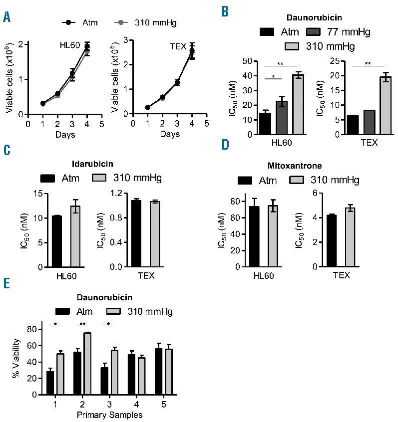
Figure 1.

Acute myeloid leukemia (AML) cells at increased pressure display chemoresistance to danorubicin. (A). Cell viability measured by trypan blue exclusion assay of HL60 and TEX leukemia cells cultured over 4 days at 37°C at atmospheric pressure (atm) and 310 mmHg above atm. (B). HL60 and TEX cells were cultured at 37°C at atm and 77 mmHg and 310 mmHg above atm for three days, and then treated with increasing concentrations of daunorubicin at indicated pressures for another three days. Cell viability was determined by Celltiter Fluor assay. *P<0.05 and **P<0.0001 as determined by one-way ANOVA with Bonferroni post-test. (C and D). HL60 and TEX cells were cultured at 37°C at atm and 310 mmHg above atm for three days, and then treated with increasing concentrations of idarubicin (D) and mitoxantrone (E) at the same pressures for another three days. Cell viability was determined by Celltiter Fluor assay and IC50 were calculated. (E) Patient samples were cultured at 37°C at atm and 310 mmHg above atm for three days, and then treated with 250 nM daunorubicin for three days at the same pressure levels. Cell viability was determined by Celltiter Fluor assay. *P<0.01 and **P<0.001 as determined by Student t-test. In all panels, data represent the mean ± standard deviation of three independent experiments.
