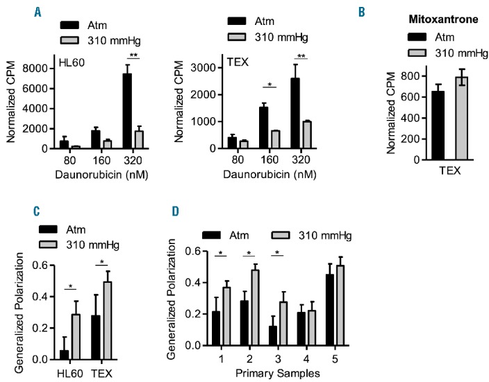
Figure 2.

AML cells have reduced daunorubicin accumulation at increased pressure. (A). HL60 and TEX cells were cultured at 37°C at atm and 310 mmHg above atm for 3 days and then treated with 80, 160 and 320 nM of [3H] daunorubicin for 3 hours at the same pressure levels. Following treatment, intracellular levels of radiolabeled daunorubicin were measured using a scintillation counter. *P<0.01 and **P<0.0001 as determined by two-way ANOVA with Bonferroni post-tests. Mean ± SD, n=3. (B) TEX cells were cultured at 37°C at atm and 310 mmHg above atm for 3 days and then treated with 8 nM of [3H] mitoxantrone for 3 hours at the same pressure levels. Following treatment, intracellular levels of radiolabeled mitoxantrone were measured using a scintillation counter. Mean ± SD, n=3. (C–D). HL60, TEX, and primary AML patient cells (1 × 10⁶ cells) were cultured at 37°C at atm and 310 mmHg above atm for 3 days, and then stained with 10 μM of the lipophilic probe, laurdan for 30 minutes. The emission state of laurdan was assessed using two-photon microscopy at 37°C, atm and within 10 minutes of removing from the pressure chamber. The fluorescence spectrum of laurdan is sensitive to the physical state of membrane phospholipids; therefore, when excited, laurdan emits at 440 nm when the membrane is in a gel-like state and at 490 nm when the membrane is in a liquid-crystalline phase. The generalized polarization (GP) factor was calculated as described in the Supplemental data. *P<0.0001 as determined by Student’s t-test. Data represent the mean ± SD of GP values from 30–60 cells.
