FIGURE 2.
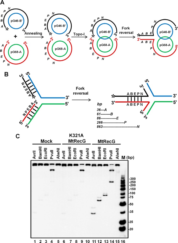
M. tuberculosis RecG catalyzes efficient fork reversal with plasmid-based replication fork structure. A, schematic representation of assembly of plasmid-based replication fork structure. Annealing of the 5′-phosphate-labeled pG46-B with pG68-A followed by topoisomerase I treatment results in catenation of the two plasmids through the plectonemic joint. B, schematic diagram of RecG-mediated fork reversal of the substrate shown in A. Fork reversal activity leads to annealing of the two strands (regressed arm) that contain restriction endonuclease sites (A, AvrII; B, BamHI; E, EcoRI; P, PvuII and N, AlwnI). Expected products of indicated restriction endonucleases are shown below. C, analysis of fork reversal products with wild-type MtRecG and ATPase-deficient MtRecG. Reaction mixture contained 2 nm replication fork substrate in a reaction buffer containing 1 mm ATP and 5 mm MgCl2 in the absence (lanes 1–5) or presence of either 10 nm K321A MtRecG (lanes 6–10) or presence of 10 nm MtRecG (lanes 11–15). Reactions were stopped with addition of ATPγS and subsequently digested with indicated restriction enzymes. Products were resolved on 10% native polyacrylamide gel and visualized by autoradiography. Lane 16, ultra-low range (10–300 bp) DNA ladder.

