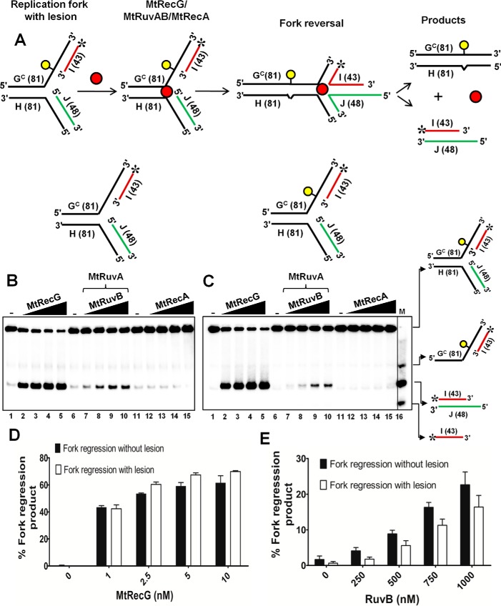
FIGURE 9.
M. tuberculosis RecG but not RuvAB promotes efficient reversal of fork structures that contain a lesion on the leading strand template DNA. A, schematic representation of a homologous replication fork containing iso-C lesion on the leading strand template and the outcome of RFR. The asterisk indicates 32P label. Reaction mixture contained 10 nm either normal homologous replication fork substrate (B) or replication fork with an iso-C lesion on the leading strand template DNA (C) in a reaction buffer containing 1 mm ATP and 5 mm MgCl2 in the absence (lanes 1, 6, and 11) or presence of 1, 2.5, 5, and 10 nm MtRecG (lanes 2–5, respectively), 250 nm MtRuvA with 250, 500, 750, and 1000 nm MtRuvB (lanes 7–10, respectively), and 125, 250, 500, and 1000 nm MtRecA (lanes 12–15, respectively). Lane 16 represents marker. Quantitative data for the MtRecG- (D) and MtRuvAB (E)-mediated fork reversal of substrates with and without template lesion. The error bars represent standard error of the mean (S.E.) from three independent experiments.

