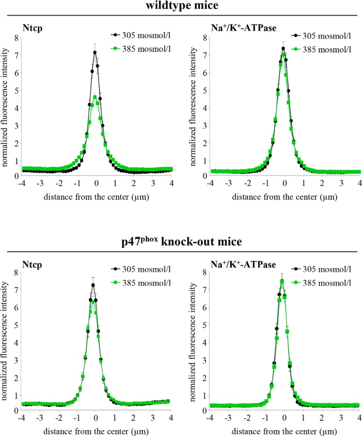
FIGURE 4.
Distribution of Ntcp in hyperosmotically perfused wild-type and p47phox knock-out mouse liver. Mouse livers were perfused for 30 min with either normo-osmotic (305 mosmol/liter, black) or hyperosmotic Krebs-Henseleit buffer (385 mosmol/liter, green) and stained for Ntcp and Na+/K+-ATPase. Densitometric analysis of fluorescence profiles and intensity of Ntcp and Na+/K+-ATPase staining is shown. The means ± S.E. of 30 measurements in each of three individual experiments for each condition are shown. As shown by colocalization with Na+/K+-ATPase, Ntcp is largely localized in the membrane under normo-osmotic conditions. After hyperosmotic perfusion, Ntcp is no longer colocalized (p < 0.05) with Na+/K+-ATPase and appears inside the cells in wild-type animals but remained unchanged in p47phox knock-out mice.

