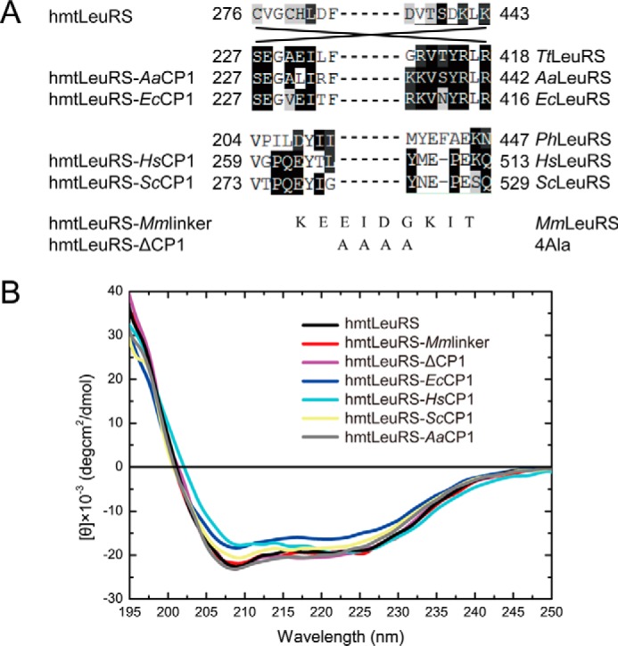
FIGURE 2.

Construction of clones encoding mosaic enzymes and CD spectroscopic analysis of these proteins. A, schematic demonstration of detailed fusion sites of the chimeric proteins. The definition of CP1 domain was based on the crystal structure of TtLeuRS (PDB code 2BTE) and PhLeuRS (PDB code 1WKB) and sequence alignment. Numbers represent the beginning and end of each CP1 domain in the context of full-length enzyme. The abbreviations are the same as Fig. 1, except for two species (Ph, P. horikoshii; Mm, M. mobile). B, CD spectra suggesting the proper secondary structure of hmtLeuRS and its derived mosaic enzymes.
