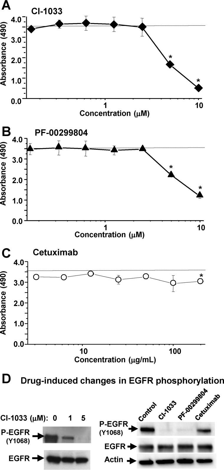
FIGURE 3.
Responses of EGFR-driven cancer cells to EGFR inhibitors. A–C, A431 cells harboring genomic amplification of the EGFR gene were grown in monolayer cultures and exposed for 24 h to the indicated irreversible EGFR kinase inhibitors (EKIs: CI-1033 and PF-00299804) or neutralizing anti-EGFR antibody (Cetuximab) at increasing concentrations. The cells were tested for metabolic activity using the MTS assay. EKIs, but not Cetuximab, triggered marked and dose-dependent reduction in metabolic activity. D, effects of drug treatment on EGFR phosphorylation (P-EGFR; Western blotting); numerical values represent mean ± S.D. of several independent experiments; *, p > 0.05.

