FIGURE 2.

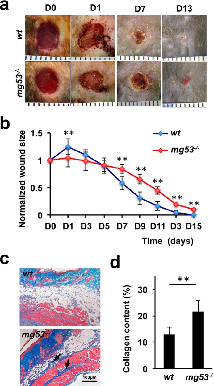
mg53−/− mice display delayed wound healing and excessive collagen deposition following injury. a, representative images of cutaneous wounds from WT (top panel) and mg53−/− (bottom panel) mice at different time points. D, day. b, statistical analyses revealed that mg53−/− mice displayed delayed wound healing compared with their WT littermates. **, p < 0.01 (n = 12). c, Masson trichrome staining of wound sections derived from WT (top panel) and mg53−/− mice (bottom panel) 10 days post-injury. Arrows indicate fibrosis and deposition of collagen. d, quantification of collagen deposition in dorsal skin on day 10 following excisional wounding in WT and mg53−/− mice. **, p < 0.01 (n = 6/group).
