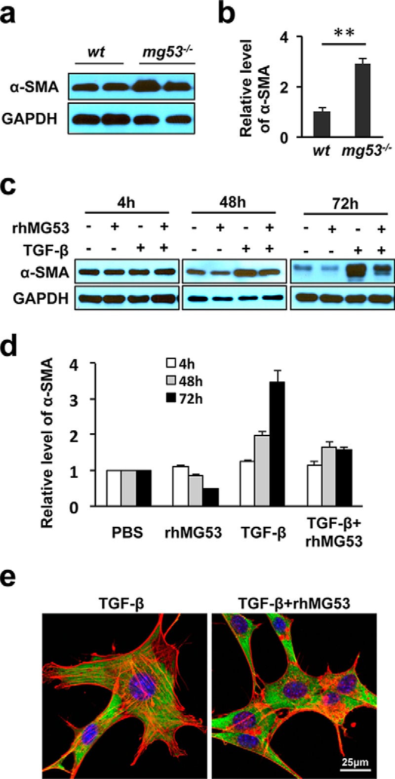
FIGURE 7.

rhMG53 treatment suppresses TGFβ-mediated α-SMA activation in fibroblasts. a, Western blotting shows elevated levels of α-SMA in skin tissues derived from mg53−/− mice compared with those from WT littermates. GAPDH was used as a loading control. b, summary data from multiple animals. **, p < 0.01. (n = 6/group). c, immunoblots of α-SMA expression in cultured 3T3 fibroblasts grown in serum-free medium in the presence of TGF-β (10 ng/ml), rhMG53 (60 μg/ml), or both TGF-β and rhMG53. Western blotting shows that rhMG53 suppressed TGF-β-mediated activation of α-SMA, with a maximal effect at the 72-h time point. d, the relative expression of α-SMA at different time points. Data are the average of three independent experiments. **, p < 0.01. e, immunofluorescence staining of α-SMA (green) and F-actin (phalloidin staining, red) reveals the reduction of stress fibers in cells treated with TGF-β plus rhMG53 (right panel) compared with cells treated with TGF-β alone (left panel) (n = 3 independent experiments).
