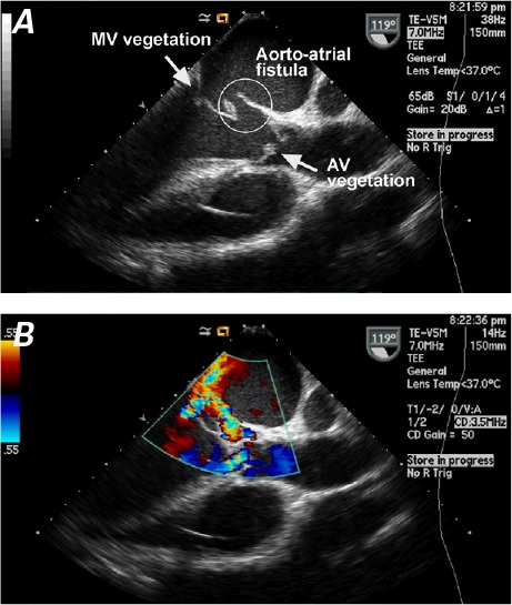
Fig. 2.

Two-dimensional transesophageal echocardiogram shows A) an aorto–left atrial fistula, together with vegetations on the mitral valve (MV) and aortic valve (AV). B) Color-flow Doppler mode shows the flow across the fistula, toward the left atrium.
