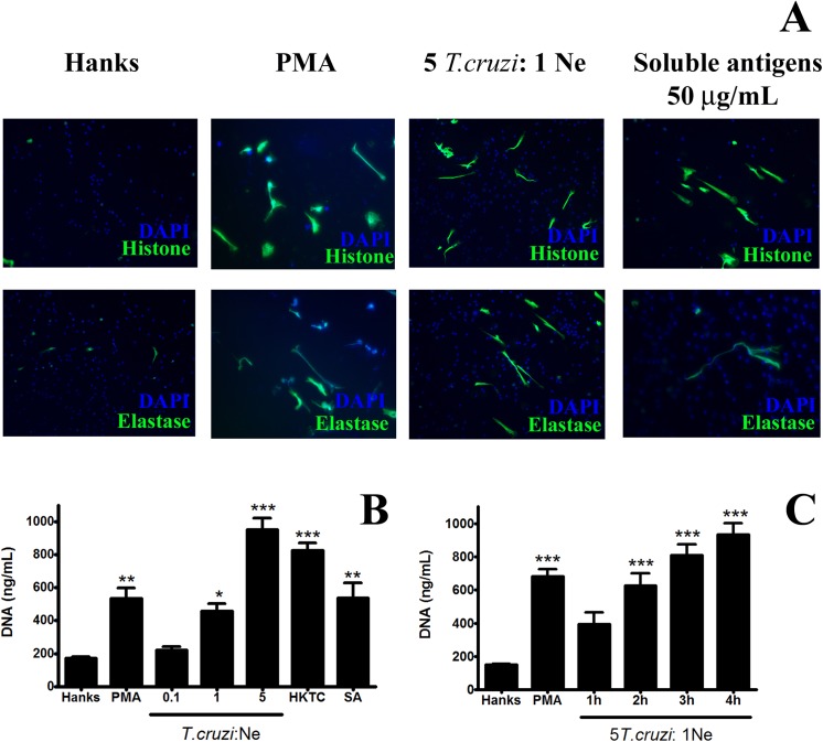
Fig 1. Trypanosoma cruzi parasites and its soluble antigens induce NET release.
Neutrophils (2 × 10⁵) were incubated with trypomastigote forms or their soluble antigens for 1–4 h and analyzed for NETs release by fluorescence microscopy and extracellular DNA quantification. (A) Neutrophils were incubated with T. cruzi (5 Tc: 1 Ne), soluble antigen (50 μg/mL), PMA (25 nM), or only HANKS for 4 h. NETs were observed by fluorescence staining using antibodies: anti-histone (green), anti-elastase (green), and fluorescein isothiocyanate-conjugated antibody and DAPI (blue). Stimulated neutrophils showed 5 ± 4 NETs by field (40× objective). (B) Neutrophils were incubated with T. cruzi in different ratios (0.1–5 parasites: 1 Ne), heat-killed T. cruzi (5 parasites: 1 Ne), and soluble antigen (50 μg/mL) for 4 h. HANKS and PMA (25 nM) were used as negative and positive controls, respectively. DNA in the supernatants was quantified using a dsDNA High Sensibility Assay Kit. (C) Neutrophils were incubated with T. cruzi (5 Tc: 1 Ne) for different time periods (1–4 h). HANKS and PMA (25 nM) were used as negative and positive controls, respectively. DNA in the supernatants were quantified as described in B. All experiments were conducted in triplicate with at least 3 independent assays. The results (B, C) were analyzed by ANOVA followed by Bonferroni multiple comparisons test. Asterisks indicates significant differences when compared with the control group (HANKS) (*P < 0.05, **P < 0.01, ***P < 0.001).

