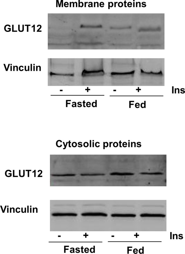
Fig 8. GLUT12 translocation after insulin stimulation.

In the upper part, a representative Western blot shows GLUT12 content in membrane fractions prepared from leg muscle of fasted and fed animals without insulin injection or 1hr after insulin injection (1U/kg). In the lower part, a representative Western blot shows GLUT12 content in the corresponding cytosolic proteins used as a control of the cytosolic GLUT12 content. Vinculin was used as a loading control for the two fractions.
