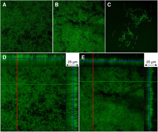
Fig 3. Confocal laser scanning microscopy analysis of rodent Pasteurellaceae biofilms.

The type reference strains of the three rodent Pasteurellaceae species studied were allowed to produce biofilms for 24 h on glass coverslips and then examined by CLSM as described in Materials and Methods. The upper panel images are two-dimensional images of the biofilms formed by [P.] pneumotropica biotype Jawetz (A), [P.] pneumotropica biotype Heyl (B) and [A.] muris (C). The lower panels are orthogonal views of z-stacks of [P.] pneumotropica biotype Jawetz (D) and [P.] pneumotropica biotype Heyl biofilms (E) where the larger panel is a “bird´s eye” view of the biofilms whereas the right and the upper panels are side views of x- and y-axis sections respectively.
