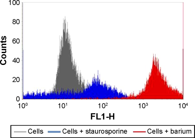
Figure 2.

Apoptosis of colostral MN phagocytes exposed to barium (1 ng/nL) and staurosporine (Sigma-Aldrich Co., St Louis, MO, USA) indicated by fluorescence intensity.
Notes: Cells were stained with Annexin V-FITC (Sigma-Aldrich Co.). Immunofluorescence analyses were carried out by flow cytometry (FACScalibur; BD Biosciences, San Jose, CA, USA).
