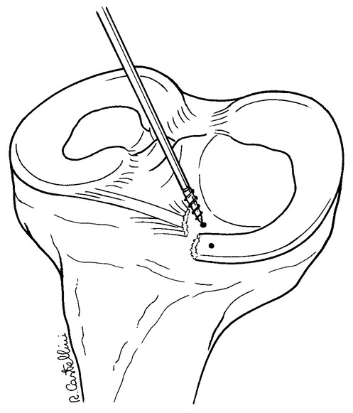
Figure 3–4.


Remnant tissue are removed at the root attachment site. The anterior horn of the meniscus is carefully reduced with a 19 gauge spinal needle, and then with an arthroscopic grasper, assessing quality and tension of the meniscus at the new insertion site.
