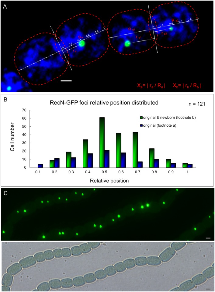
Fig 3. Segregation of RecN foci during vegetative cell division.
(A) The method used to quantify the relative distance between foci along the cell division axis. In the formula, “a” means a cell inheriting the original RecN focus after division; “b” means a cell with both an original and a newborn RecN focus. Photographs were captured by using an Olympus FV1000 confocal microscope. Cells were stained with DAPI (blue). Scale bars correspond to 1 μm. (B) Cell number distribution obtained according to the relative distance. (C) Original and newborn RecN foci usually were nearly symmetrically distributed along the cell division axis. Photograph by using a Nikon Eclipse 80i microscope, in the fluorescence and bright fields. Scale bars correspond to 1 μm.

