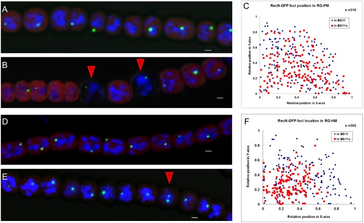
Fig 6. RecN localization is affected in patS or hetR mutant.
(A) RecN-GFP foci in the strain RG-PM cultured in the medium BG11. (B) RecN-GFP foci in the strain RG-PM cultured in the medium BG110. The red arrows indicate heterocysts. (C) The localization of RecN-GFP foci in the strain RG-PM cultured in different media. The coordinate 0 is the center of the cell. The statistical method used here was the same with that in Fig 1B. (D) RecN-GFP foci in the strain RG-HM cultured in the medium BG11. (E) RecN-GFP foci in RG-HM cultured in the medium BG110, the red arrow indicates a cell with 2 foci. (F) The localization of RecN-GFP foci in the strain RG-HM cultured in different media. The coordinate 0 is the center of the cell. The statistical method used here was the same with that in Fig 1B. Photographs were taken by using an Olympus FV1000 confocal microscope. Cells were stained with DAPI (blue). The red fluorescence is from the photosynthetic pigments. Scale bars correspond to 1 μm.

