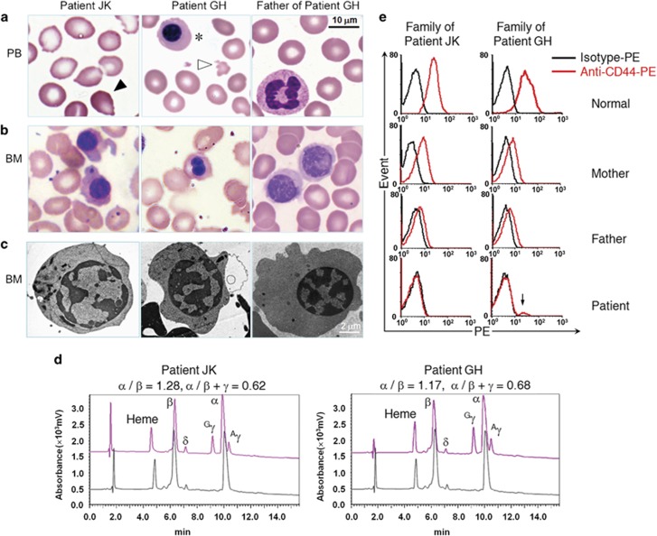
Figure 1.
Phenotypic analyses of the patients in this study. (a) Peripheral blood smears from patients JK and GH show tear drop-shaped poikilocytes (black arrow), microcytosis and hypochromasia, fragmented erythrocytes (white arrow), and nucleated erythroblasts (asterisks). (b) Bone marrow smears show poikiloblast. (c) TEM analysis shows erythroblasts with heterochromatin clumps around the nuclear periphery. (d) Reversed-phase high-performance liquid chromatographic analysis of globin chains in JK, GH, and a healthy individual. Peaks and corresponding retention times for heme groups and different globin chains are indicated (red, patient; black, control). Data for α/β and α/β+γ ratios are shown above the chromatograms. (e) Flow cytometric analysis of CD44 expression in mature erythrocytes shows erythroid-specific CD44 deficiency in patients and reduced expression in their heterozygous parents. The mixed phenotype with low CD44 expression (arrow) likely arose from still-circulating transfused blood (50 days after the last transfusion).

