Figure 4.
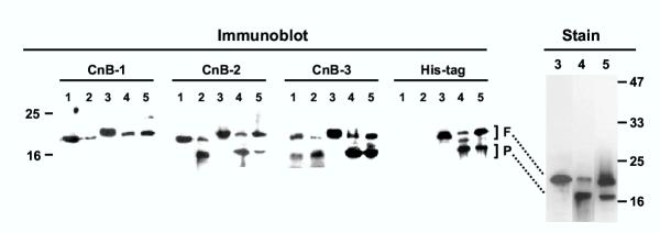
Processing of PfCnB in vivo and in vitro. Immunoblot analysis was performed with the antibodies CnB-1, -2 and -3 (described in Fig. 2) and the anti-His tag antibody on the following samples. 1 = total protein (60 μg) from freshly isolated parasite; 2 = same as lane 1 except that the parasite was incubated at room temperature for 5 min; 3 = purified recombinant His-tagged PfCnB (120 ng); 4 = as in lane 3 but after incubation with parasite extract; 5 = as in lane 4 except that the incubation mixture also contained protease inhibitor cocktail. In the right panel (Stain) higher quantities (5 μg) of the same recombinant protein samples of the corresponding lane numbers were analyzed and the gel stained with Coomassie Blue R250. The full-length and processed bands are indicated as F and P, respectively. Size markers are indicated on the two sides. Details are given in Methods.

