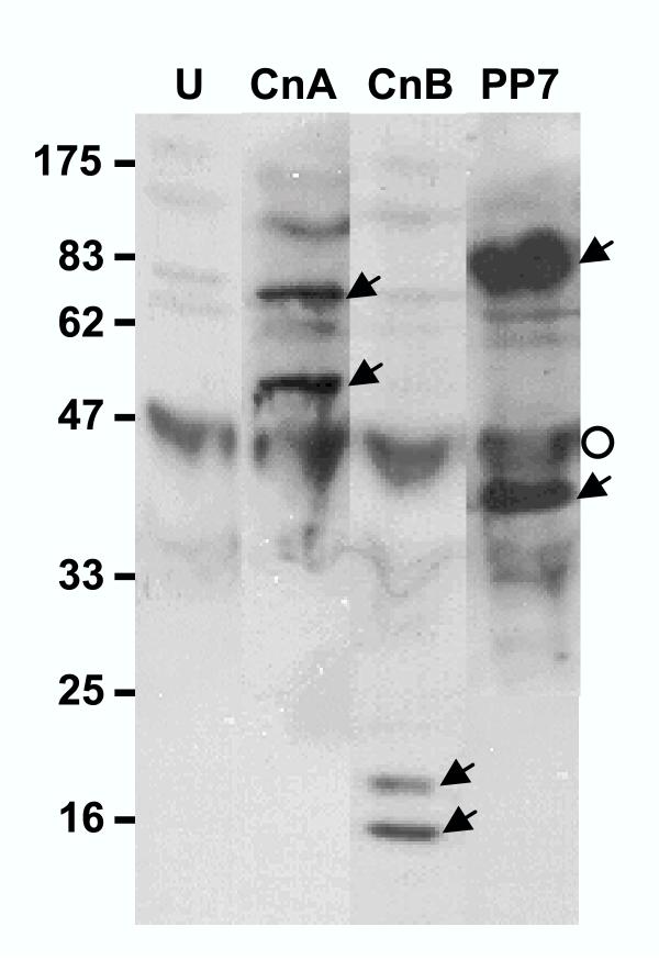
Figure 5.
Processed phosphatases in vivo. P. falciparum-infected erythrocytes (RBC) at 15% parasitemia were centrifuged at 12,000 × g in cold for 1 min and the pellet resuspended in 50 μl SDS sample buffer [32], followed by boiling (5 min). The undissolved material (mostly RBC skeleton and parasitic hemozoin) was removed by centrifugation and the supernatant (250 μg protein per lane) analyzed by immunoblot using internal peptide antibodies against the indicated proteins (CnA-2, CnB-2 and PP7-2, respectively; compare with the bands in Fig. 3, 4, and 7, respectively). The two bands (full-length and processed) in each lane are indicated by arrowheads. Lane U represents a similarly processed sample from uninfected RBC, probed with a 1:1:1 mixture of all three antibodies. The background bands are due to nonspecific binding of the IgG's to various RBC components, the darkest of which is marked by the open circle. The size markers are shown on the left.

