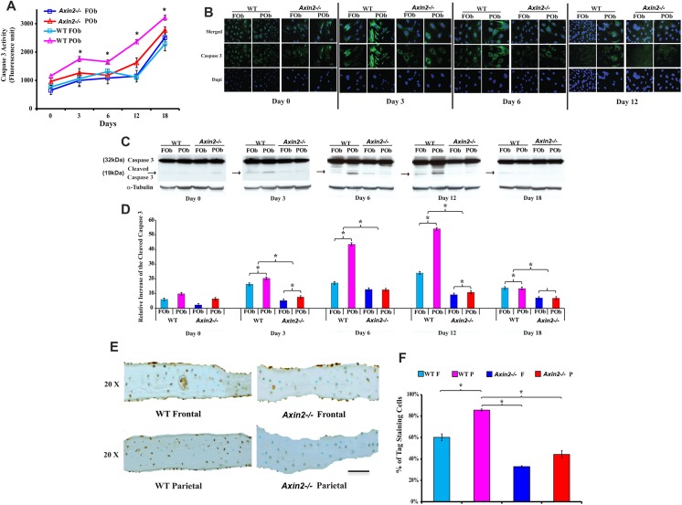
Fig 3. Decreased apoptotic activity in Axin2 -/- FOb and POb and calvarial bones.
(A) Apoptotic activity in Axin2 -/- FOb and POb undergoing to differentiation measured as Caspase 3 fluorescence units. Axin2 -/- FOb and POb cells show similar level of Caspase 3 activity, and wild type POb the highest level. Apoptosis increased over time during the osteoblast differentiation. (B) Caspase 3 indirect immunofluorescence analysis showing intense nuclear and cytoplasmic fluorescence in wild type POb at day 3 and 6 of osteogenic differentiation, whereas only faint cytoplasmic staining is observed in Axin2 -/- POb. (C) Detection of active cleaved Caspase 3 by immunoblotting analysis. A 19 kDa band corresponding to cleaved Caspase 3 is detected in wild type POb starting from day 3, of osteogenic differentiation. (D) Histograms representing quantification of each electrophoretic band obtained by Image J program. Each band was normalized to α-tubulin loading control. (E) TUNEL staining performed on coronal sections derived from Axin2 -/- and wild type parietal and frontal bones harvested from pN21 mice. (F) Quantification of the Caspase 3 activity in bones by calculating the percentage of the positive Tag stained cells (brown stain) over the total cells in similar areas of the bone shows the highest apoptotic activity in parietal bone of wild type mice, while apoptotic activity in parietal bone of Axin2 -/-mice is significantly reduced. Scale bar = 150 μm.

