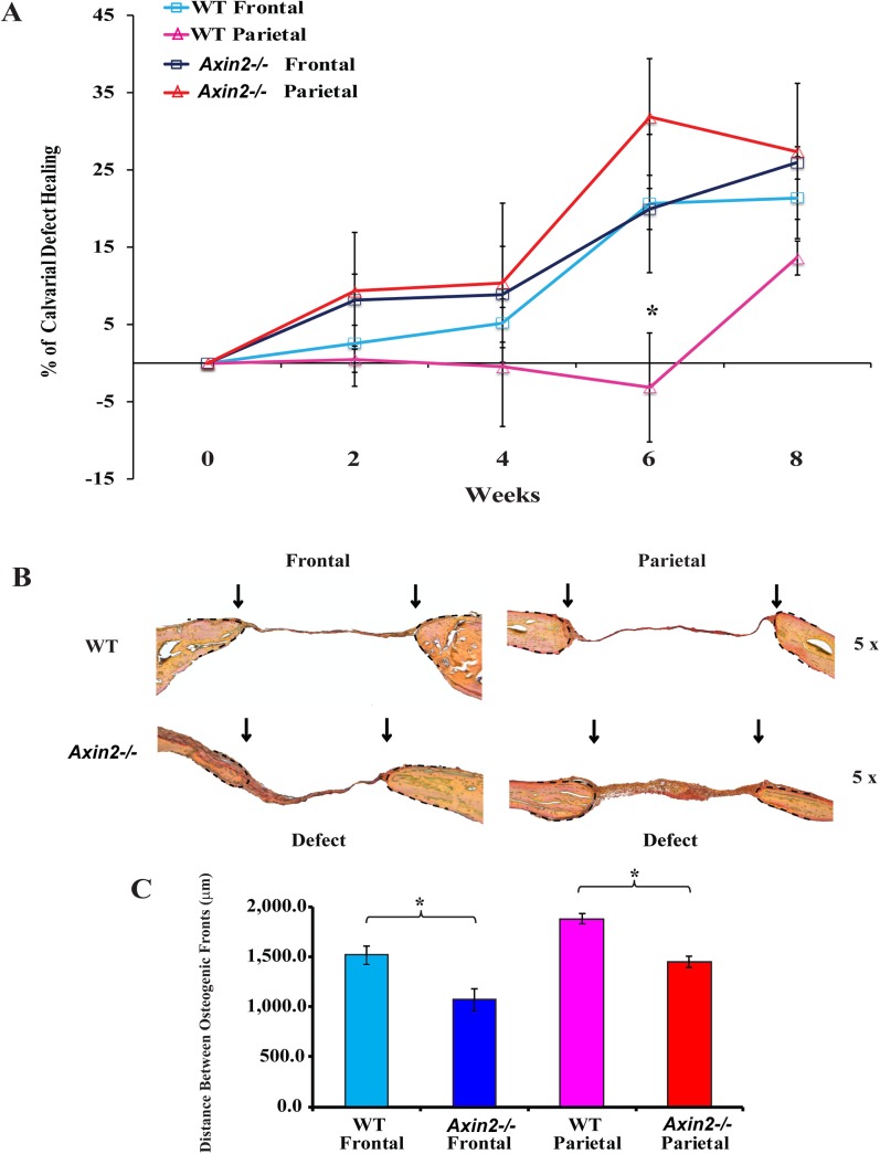
Fig 4. In vivo calvarial healing of Axin2 -/- and wild type frontal and parietal bones.
(A) Two-millimeter (2mm) defects were created in the frontal and parietal bones of 7 month-old Axin2 -/- and wild type mice (n = 3). Quantification of defect repair according to microCT-scan results. Statistical analysis was conducted utilizing the Mann-Whitney Test. P-values: *P≤ 0.05. (B) Pentachrome staining of coronal sections of skull at post-operative week 8 showing the repair of calvarial bone defects as determined by yellow color. Bone regeneration was higher in Axin2 -/- frontal and parietal bones as compared to wild type bones. (C) Histogram showing the distance between the osteogenic fronts (dashed) and marked by arrows (Objective magnification 5x).

